

")
Research activity
The cornea is covered by a thin layer of tear film that plays a protective and nutritive role, and creates optically smooth surface. Traditional clinical methods for assessing tear film surface quality are often invasive, subjective and unreliable. For several years, I have been involved in the development of new promising techniques for noninvasive objective assessment of tear film surface quality.
The main research projects focus on the following problems:
„The analysis of post-blink tear film surface quality. Understanding eatiologies of ocular surface diseases.” (Project supported by the National Science Centre) |
|
| The tear film that covers the anterior corneal surface has a dynamic behavior. It is well established that immediately after a blink, the tear film undergoes formation (build up) that is followed by a phase of relative tear film stability. However, if the eye is left open for a sufficiently long period of time, the tear film will deteriorate and break-ups in tear film will appear. For those diagnosed with a dry eye syndrome the tear film break-ups appear much sooner. Lateral shearing interferometry (LSI) has been used to objectively and quantitatively assess the tear film surface quality in a noninvasive manner of normal and dry eye subjects. Using a series of cases, we showed that LSI equipped with a set of robust parameter estimation techniques was able to characterize up to five different phases of tear film surface kinetics that include: (i) initial fast tear film build-up phase, (ii) further slower tear film build-up phase, (iii) tear film stability, (iv) tear film thinning, and (v), after a detected break-up, subsequent tear film deterioration (see figure). The two-step tear film build-up process is a subject of our current study. We aim to develop new LSI technology with focus on post-blink changes in tear film surface quality and determine key physical parameters of this phase, that may carry important information regarding tear film quality and its behaviour across the whole inter blink interval. |
|
More information in: Szczesna-Iskander, TOS 2017 Szczesna & Iskander, OVS 2010 Szczesna & Kasprzak, OPO 2009 |
|

|
|
|
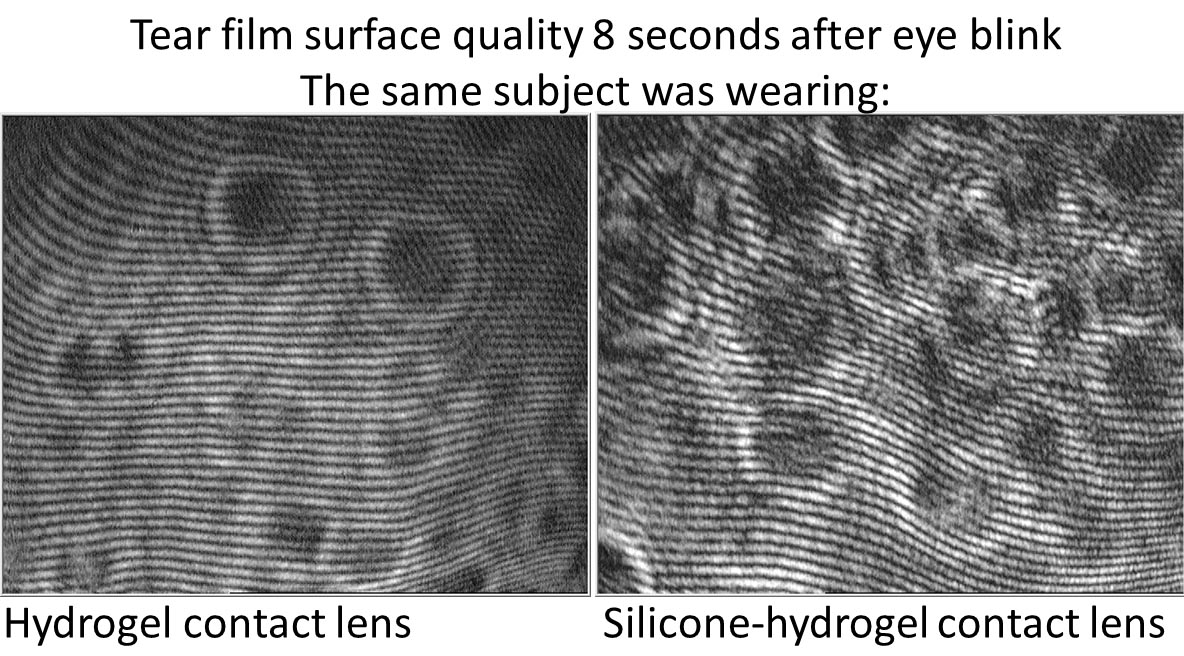
| Contact lens–induced tear film changes are significant clinically because a large proportion of contact lens wearers report dry eye symptoms, and symptoms of dryness have been found to be a primary reason for contact lens intolerance. Lubricants like artificial tears provide only symptomatic relief. There is a great selection of contact lens materials, designs and lubricating drops. An advanced diagnostic system based on high-speed videokeratoscopy, lateral shearing interferometry and dynamic OCT based meniscometry is being developed for non-invasive in vivo assessment of tear film quality. The dynamic behaviour of tear film on contact lens surface will be quantitatively described by a set of robust estimators to estimate clinically relevant characteristics of the tear film. It is expected that the new algorithms will bring consistency and repeatability to the results of tear quality assessment making future diagnosis and monitoring of contact lens induced dry eye more reliable and provide the opportunity to assess the effect of contact lens materials on eye as well as the effect of artificial tears. |
|
More information in: Szczesna-Iskander et al., OVS 2016 Szczesna-Iskander et al., OVS 2014 Szczesna-Iskander et al., IOVS 2012 Szczesna, ECL 2011  |
|
“Predicting Dry Eye Using Noninvasive Techniques of Tear Film Surface Assessment” (Research project partially supported by Endeavour Fellowship in collaboration with Contact Lens and Visual Optics Laboratory, Queensland University of Technology, Australia) |
||
| There are many well-established techniques and tests for diagnosing dry eye. However, the reliability of many of the traditional clinical tests for dry eye diagnosis has been brought into question. We applied three noninvasive techniques for measurement of tear film surface quality: dynamic-area high-speed videokeratoscopy (HSV), wavefront sensing (DWS), and lateral shearing interferometry (LSI). Our aim was to establish the ability of these noninvasive techniques to predict dry eye. The tear film surface quality was measured in healthy and dry eye subjects. Results from those methods were compared to standard clinical procedures based on slit lamp examination. LSI technique showed the best detection performance, closely followed by the dynamic-area HSV. Such an assay was achieved in natural blinking conditions as well as suppressed blinking conditions (see ROC (Receiver Operating Characteristics) curves: comparison between, natural blinking conditions (NBC) and suppressed blinking conditions (SBC) of those two methods). Importantly, this project showed that noncontact techniques for assessing tear film surface quality in truly noninvasive measurement conditions (natural blinking conditions) have considerable potential for the objective detection of dry eye. | 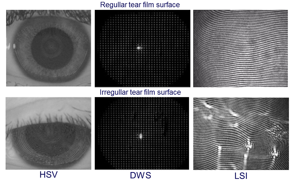 |
|
| More Information in: Szczesna et al., IOVS 2011 Szczesna et al., JBO 2010 |
|
|

“Examination of precorneal tear film after refractive surgeries e.g. LASIK, RK” (Project was conducted in collaboration with Prof. Ulf Stenevi, Sahlgren’s University Hospital, Mölndal, Sweden)
|
|
| The popularity of refractive surgery has grown exponentially over the last two decades. It is well known that refractive surgeries can cause dry eye symptoms—unstable tear film, which is associated with morphological and physiological changes in ocular surface and the tear function. The lateral shearing interferometry was used for measurement of tear film on post LASIK (laser in situ keratomileusis) and RK (radial keratotomy) eyes. Image processing procedures were used to analyse the observed pattern of tear film breakups (tear film breakups appear as high-intensity patterns bright lines in the background of the interferogram). The numerical analysis of recorded images revealed the repeatability of breakup patterns. It might suggest that tear film breakup may be associated with local irregularities of the corneal topography arisen as a consequence of LASIK and RK surgeries. | 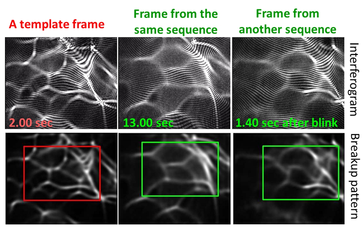 |
| More information in: Szczesna et al., J Biomed Opt. 14(6): 064029, 2009 |
|